
The human vascular system responds with remarkable sensitivity to the repetitive, low-impact mechanical stimulus of walking. While vigorous exercise often receives greater attention in clinical literature, sustained ambulation at modest intensities initiates a distinct sequence of hemodynamic adaptations. When an individual walks, rhythmic skeletal muscle contractions in the lower limbs compress intramuscular veins, periodically increasing venous return to the heart and prompting an acute rise in cardiac stroke volume. Over time, these daily mechanical shifts alter endothelial signaling, modify arterial compliance, and relieve systemic vascular resistance.
For individuals seeking to sustain or restore cardiovascular function, walking presents an accessible yet scientifically robust intervention. It imposes minimal orthopedic strain while delivering sufficient volume to preserve capillary density and arterial elasticity throughout the peripheral vasculature. To comprehend the complete scope of this systemic shift, one must examine the specific mechanics within the vessel walls, establish reliable measures for baseline effort, and observe how modest structural variations in duration, frequency, and terrain guide long-term circulatory adaptations.
Vascular Mechanics Under Sustained Low-Intensity Load
At the microscopic level, sustained walking introduces steady laminar blood flow across the inner lining of the arterial tree. This fluid motion produces shear stress against endothelial cells, triggering the enzymatic activity of endothelial nitric oxide synthase. The resultant release of nitric oxide diffuses into the adjacent vascular smooth muscle layer, prompting cyclic guanosine monophosphate synthesis and subsequent vasodilation. Consequently, peripheral vascular resistance declines during the walk, allowing blood to circulate with reduced downstream opposition.
This localized reduction in vascular resistance is accompanied by the active engagement of the peripheral venous pump. With each heel-strike and subsequent toe-off, the soleus and gastrocnemius muscles contract against the deep fascia, compressing the posterior tibial, peroneal, and popliteal veins. One-way intraluminal valves prevent retrograde flow, propelling venous blood toward the inferior vena cava and right atrium. This process sustains ventricular preload without requiring an excessive surge in sympathetically driven heart rate, contrasting sharply with the cardiovascular dynamics of higher-intensity sprinting.
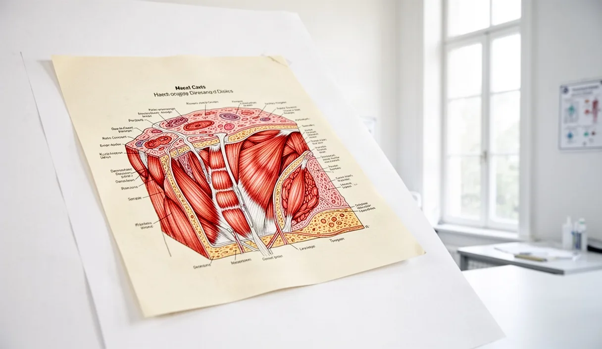
Over a prolonged period, regular exposure to this rhythmic mechanical stimulus preserves vascular compliance. Arterial stiffness, often accelerated by sedentary habits and advanced chronological age, is characterized by the breakdown of elastin fibers and an accumulation of rigid collagen within the tunica media. Daily walking counteracts this matrix remodeling. Regular mechanical distension preserves the functional architecture of elastic arteries, such as the aorta and carotid vessels, which helps to attenuate excessive pulse pressure waves before they reach vulnerable microvascular beds in the kidneys and brain.
Furthermore, daily low-intensity ambulation promotes microvascular maintenance within skeletal muscle tissue. As metabolic demand persists over thirty to sixty minutes, capillary beds that remain dormant during seated rest are systematically recruited. This capillary perfusion enhances oxygen extraction efficiency at the tissue level, permitting cardiac output to serve active metabolic needs with greater mechanical economy.
Assessing Your Baseline Walking Cadence
To implement walking as an intentional physiological practice, one must establish an accurate measure of ambulation speed and cadence. Cadence, defined as the number of steps taken per minute, provides an objective metric that correlates directly with metabolic cost, vertical ground reaction force, and cardiovascular demand. Relying solely on distance can introduce ambiguity, as stride length varies considerably across individuals based on height, limb geometry, and joint mobility.
Determining your current baseline requires a standardized observation period rather than an estimation. You may measure this by identifying a flat, unobstructed pedestrian path and following a clear diagnostic protocol:
- Fasten a comfortable pair of walking shoes and select an accurate timing device or wrist-worn accelerometer.
- Walk at your normal, unhurried pace for five minutes to reach a steady circulatory state.
- Once this initial interval concludes, start a sixty-second timer.
- Count every step taken by both feet during that full sixty-second window, or record the total registered by an accurate sensor.
- Repeat this measurement on three distinct days at approximately the same time of day, calculating the arithmetic mean of the three results.
The resulting figure establishes your spontaneous cadence. In adult populations, spontaneous walking cadence typically falls into three primary operational bands, each representing a distinct degree of cardiovascular engagement:
| Cadence Band | Steps Per Minute | Physiological Focus | Primary Circulatory Influence |
|---|---|---|---|
| Slow / Base | 70 to 89 | Postural stabilization and joint mobilization | Gentle venous return, minimal systemic pressor response |
| Moderate Maintenance | 90 to 114 | Aerobic base and continuous capillary flow | Steady shear-mediated nitric oxide synthesis, mild stroke volume expansion |
| Brisk / Structured | 115 to 134 | Cardiovascular conditioning | Noticeable rise in cardiac output, enhanced peripheral oxygen extraction |
Once you identify your primary bracket, modifications should be made conservatively. If your spontaneous cadence is 84 steps per minute, an appropriate target for structured sessions is 92 to 96 steps per minute, rather than an abrupt transition to a fast rate that might destabilize gait mechanics or strain connective tissues.
Continuous Versus Segmented Walking Intervals
A frequent subject of clinical and practical inquiry is whether walking yields greater cardiovascular benefit when conducted as a solitary, prolonged bout or when divided into multiple brief intervals across the day. The answer depends largely on the specific cardiovascular and metabolic markers under consideration, as continuous and segmented walking stimulate circulatory mechanics in slightly different manners.
Continuous walking, performed for thirty to sixty uninterrupted minutes, provides a sustained hemodynamic steady state. During an extended session, core body temperature increases slightly, promoting skin blood flow and sustained peripheral vasodilation. Blood pressure often displays a marked post-exercise hypotensive response following a long session, with systolic measurements resting several millimeters of mercury below baseline for several hours. This extended stimulus also tests cardiac stroke volume over time, encouraging structural myocardial efficiency and sustained vagal modulation once the session terminates.
Segmented walking, such as three distinct twelve-minute bouts distributed throughout the day, exerts a pronounced effect on postprandial glucose dynamics and acute arterial stiffening. Walking for ten to fifteen minutes shortly following a meal engages skeletal muscle contraction when blood glucose and insulin concentrations are rising. This muscular activity stimulates GLUT4 glucose transporter translocation through insulin-independent pathways, blunting acute glycation stress and the transient endothelial dysfunction that typically follows a heavy nutritional load.
| Variable | Continuous Session (45 Minutes) | Segmented Bouts (Three 15-Minute Sessions) |
|---|---|---|
| Cardiovascular Endurance | Higher sustained stroke volume duration | Moderate, resets between sessions |
| Endothelial Shear Stress | Prolonged, unbroken exposure | Intermittent, recurrent activation |
| Postprandial Glycemic Control | Dependent on session timing relative to meals | Consistently blunts immediate glucose surges |
| Orthopedic Recovery Demand | Higher cumulative friction per bout | Lower, provides connective tissue rest |
| Post-Exercise Hypotension | Deeper, longer single hypotensive period | Multiple smaller systemic pressure drops |
For individuals managing persistent elevated blood pressure, a continuous block often produces the most consistent reduction in twenty-four-hour systemic vascular load. Conversely, for those contending with erratic blood glucose concentrations or marked sedentary stiffness from office labor, distributing walks across the day provides superior metabolic protection and frequent venous decompression.
Practical Adjustments for Variable Terrain
While walking along flat pavement provides a dependable baseline, introducing variations in surface texture and gradient alters the cardiovascular workload without requiring increases in velocity. Modifying the physical environment challenges the heart to accommodate changing muscular demands, effectively varying myocardial work while maintaining low orthopedic impact.
Ascending an incline requires immediate increases in mechanical work from the gluteal muscles, hamstrings, and calves. To overcome gravitational resistance, the motor units within these muscle groups fire at higher frequencies, demanding greater oxygen delivery. Consequently, systemic arteriolar dilation deepens in the lower extremities, prompting the heart to increase both stroke volume and myocardial contractility to sustain perfusion pressure. A grade increase of merely 3% to 6% can raise heart rate by 18 to 32 beats per minute at an identical walking velocity, transforming a gentle walk into an efficient cardiovascular conditioning stimulus.
Conversely, descending an incline emphasizes eccentric muscle contractions. During eccentric action, the quadriceps and tibialis anterior lengthen under tension to decelerate the body. While eccentric contractions generate lower systemic metabolic demand than concentric ascents, they impose unique mechanical stress that stimulates microvascular repair pathways and tendon resilience. When descending, you must shorten your stride slightly and land with a midfoot strike to prevent excessive impact transmission through the patellofemoral and hip joints.
Surface compliance also alters cardiovascular mechanics. Consider these primary outdoor substrates and their physiological characteristics:
- Asphalt and Concrete: Provide maximum mechanical energy return and reliable traction. These surfaces make cadence regulation straightforward, though they generate higher ground reaction forces that travel up the kinetic chain.
- Packed Forest Soil or Crushed Gravel: Yield slightly under the foot, absorbing a portion of the impact energy. This minor instability recruits intrinsic foot musculature and ankle stabilizers, slightly raising metabolic cost without elevating joint stress.
- Dry Sand or Deep Turf: Offer high mechanical compliance, demanding substantially greater muscular effort to maintain forward momentum. Walking on sand can increase the oxygen cost of transport by more than 40% compared to firm pavement at the same pace, placing substantial demands on the calf muscle pump and cardiac output.
When incorporating variable terrain, adjust your parameters incrementally. A sensible approach involves substituting one flat session per week with an undulating route, observing how your circulatory system tolerates the altered mechanical stress before increasing weekly exposure.
Monitoring Resting Heart Rate as an Indicator of Adaptation
The cardiovascular system adapts to regular physical training primarily by improving operational efficiency. One of the most reliable and accessible measures of this progressive adaptation is the longitudinal monitoring of resting heart rate. As daily ambulation stimulates the cardiovascular system, the balance of the autonomic nervous system gradually shifts, increasing resting parasympathetic (vagal) tone while moderating basal sympathetic tone.
This enhanced parasympathetic influence, paired with a modest increase in ventricular end-diastolic volume, allows the left ventricle to eject a greater volume of blood with each contraction. Because total cardiac output matches resting metabolic requirements, this elevated stroke volume permits the heart to beat fewer times per minute while maintaining identical systemic tissue perfusion. A steady reduction in resting heart rate over months serves as an objective marker of favorable structural and autonomic development.
To obtain reliable heart rate figures, you should adhere to a strict measuring protocol. The heart responds swiftly to emotional stress, digestion, ambient temperature, and hydration status, meaning unstandardized measurements often introduce misleading fluctuations. Follow these guidelines for accurate data collection:
- Measure your pulse immediately upon waking, before rising from bed or consuming liquids.
- Sit or lie quietly for two minutes to allow transient awakeness spikes to stabilize.
- Locate the radial artery on the thumb side of your wrist using your index and middle fingers, avoiding the use of your thumb which has its own intrinsic pulse.
- Count the beats for a full sixty seconds, or employ an accurately calibrated, validated medical device.
- Record the value in a physical notebook or secure tracking log, noting any unusual sleep disturbances or evening meals from the night prior.
Calculate your weekly average across seven consecutive days. Over an eight- to twelve-week period of regular daily walking, an individual often observes a reduction in resting heart rate of 3 to 7 beats per minute. If you observe sudden, sustained elevations in resting heart rate of 10 or more beats per minute above your baseline, this may indicate systemic fatigue, developing illness, or dehydration, signaling a need for additional recovery.
Note: If you record persistent unexplained bradycardia (below 50 beats per minute accompanied by dizziness or syncope) or sudden irregular tachycardic episodes, you should promptly consult a certified medical physician or cardiologist to evaluate your cardiac rhythm.
Common Mistakes to Avoid
Even a natural movement pattern like walking can lead to diminished returns or localized discomfort if conducted with improper technique or planning. Attention to proper execution ensures that mechanical work yields positive cardiovascular adaptation rather than musculoskeletal irritation.
- Overstriding to Increase Speed: When attempting to walk faster, many individuals reach forward excessively with the lead heel. This causes the foot to land far ahead of the body's center of mass, producing a braking force that transmits impact through the knee and hip joints while disrupting forward momentum. Instead, maintain a compact stride and increase cadence by pushing off actively with the trailing foot.
- Restricting Upper-Body Arm Swing: Holding the arms rigidly against the torso or immobilizing them with handheld weights limits the natural counter-rotation of the thoracic spine. A relaxed, reciprocal arm swing counterbalances the pelvic rotation, easing diaphragmatic breathing and supporting smooth venous blood return from the upper body.
- Neglecting Environmental Hydration and Thermal Demands: Walking in elevated heat or humidity causes blood to divert to the cutaneous vascular bed for evaporative cooling, which reduces central blood volume and accelerates heart rate drift. You must account for ambient conditions by adjusting cadence downward and consuming adequate water on warm days.
- Sudden Spikes in Weekly Volume: Transitioning from a sedentary routine to high daily mileage can overburden the plantar fascia, Achilles tendons, and hip stabilizers before vascular and structural remodeling can occur. Progress your duration by no more than 10% to 15% per week to allow connective tissues adequate time to adapt.
Practical Next Steps
Transitioning from conceptual understanding to consistent physical adaptation requires a methodical, structured strategy. Begin by establishing your current capacity rather than immediately pursuing ambitious physical milestones. For the next three days, simply carry out your standard routine while recording your baseline steps and waking resting heart rate using the protocols outlined above.
Once you have gathered this baseline data, schedule your walking sessions as fixed blocks within your weekly calendar. If you choose a segmented approach, introduce a twelve-minute walk following your largest daily meal, maintaining a cadence between 90 and 105 steps per minute. If you prefer a continuous session, designate three or four mornings per week for an uninterrupted thirty-minute walk along a level, predictable route.
Maintain this structured pattern for four consecutive weeks without altering the variables. Log your resting heart rate each morning, note any changes in physical exertion, and allow the vascular endothelium to adapt to the steady fluid shear stress. If your personal health history includes cardiovascular, metabolic, or chronic orthopedic conditions, schedule an appointment with your physician before substantially increasing your daily walking volume or altering your exercise habits. Systematic consistency, rather than acute physical strain, remains the primary driver of lasting circulatory health.