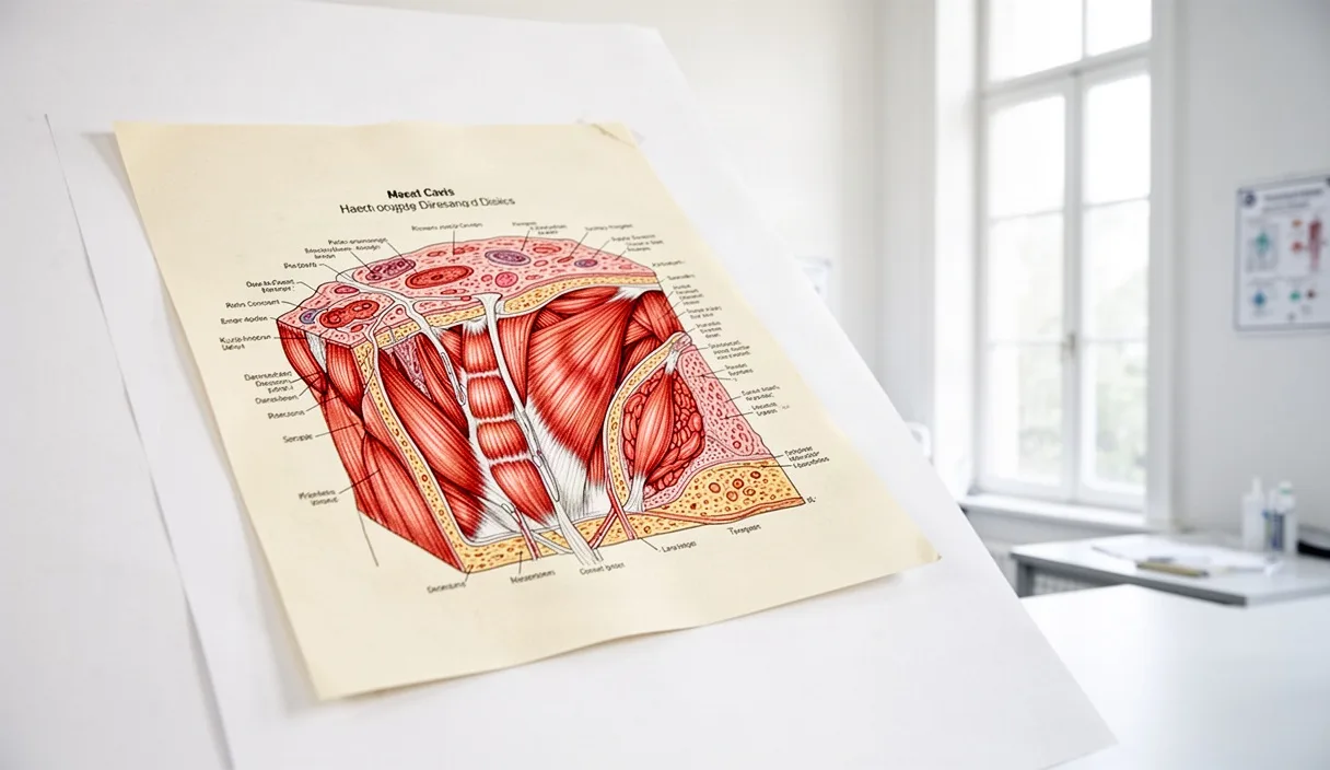
For several generations, classical anatomy textbooks characterized skeletal muscle primarily through its mechanical duties. It was classified as an organ of locomotion, posture, and heat generation, governed by electrical impulses from the central nervous system to pull against the skeletal framework. While those kinetic responsibilities remain undisputed, modern biochemical research has revealed an equally significant role: skeletal muscle serves as the primary metabolic sink in the human body, acting as an endocrine organ that communicates continuously with adipose tissue, the liver, and the vascular bed.
When muscle fibers shorten and lengthen under load, they generate humoral factors and clear energetic substrates from the bloodstream with remarkable efficiency. An adult human typically carries between 30 and 42 percent of total body mass as skeletal muscle, depending on age, biological sex, and physical conditioning. Consequently, the metabolic behavior of this single tissue dictates systemic insulin sensitivity, systemic lipid clearing, and basal energy expenditure. A failure to recognize muscle tissue as an active endocrine partner leads to incomplete approaches to metabolic health and everyday physical care.
The Endocrine Function of Contracting Muscle
The concept of skeletal muscle as an endocrine organ began to crystallize with the discovery of myokines, which are peptides synthesized and released by myocytes in direct response to contraction. Unlike standard hormones secreted by discrete glands such as the thyroid or adrenal cortex, myokines enter systemic circulation in pulses directly tied to mechanical work. Through these biochemical messengers, working skeletal muscle exerts autocrine effects on its own tissue architecture, paracrine effects on neighboring capillaries, and endocrine effects on distant viscera.
The first and most widely documented myokine is interleukin-6, commonly designated as IL-6. While systemic, chronic elevations of IL-6 from immune cells indicate chronic inflammation, muscle-derived IL-6 acts in an anti-inflammatory and metabolic fashion. When intracellular glycogen declines during sustained contraction, myocytes release IL-6 into circulation, triggering hepatic glycogenolysis to support blood glucose levels and stimulating lipolysis in visceral and subcutaneous fat depots. Concurrently, it stimulates the appearance of anti-inflammatory cytokines, including interleukin-10 and interleukin-1 receptor antagonist, while suppressing tumor necrosis factor alpha.
Other notable myokines perform distinct regulatory functions throughout the metabolic network:
- Irisin: Cleaved from the transmembrane precursor FNDC5 during sustained aerobic movement, this peptide circulates to white adipose tissue to stimulate uncoupling protein 1, encouraging mild thermogenesis and mitochondrial biogenesis.
- Myostatin: Operating largely as a negative regulator, circulating levels of this peptide decline during active resistance work, allowing myocellular hypertrophy and improving metabolic clearance capacity.
- Interleukin-15: Highly expressed in skeletal tissue, IL-15 reduces adipose mass and regulates myocellular lipid oxidation without requiring substantial muscle hypertrophy.
- Brain-Derived Neurotrophic Factor (BDNF): Although primarily studied in neural tissue, BDNF is expressed in muscle fibers upon contraction, where it enhances local fatty acid oxidation via AMP-activated protein kinase signaling.
The release of these regulatory peptides establishes that muscle contraction is not merely an energy-consuming act. Rather, it is a chemical signaling event that coordinates whole-body metabolic equilibrium across the liver, pancreas, and peripheral vasculature.
Non-Insulin Mediated Glucose Translocation
Under resting conditions, the uptake of circulating glucose into skeletal myocytes requires insulin. Following a meal, beta cells within the pancreas release insulin, which binds to cell surface receptors, initiating a cascade through insulin receptor substrate proteins and phosphoinositide 3-kinase. This pathway encourages the internal vesicles carrying Glucose Transporter Type 4 (GLUT4) proteins to move to the plasma membrane, permitting the entry of glucose into the sarcoplasm. However, during mechanical work, skeletal muscle possesses an autonomous mechanism that circumvents this requirement entirely.
When a muscle fiber contracts, intracellular calcium ions are released from the sarcoplasmic reticulum, and the ratio of cellular AMP to ATP shifts upward as high-energy phosphates are depleted. These changes activate two key enzymatic drivers: calcium/calmodulin-dependent protein kinase and 5-AMP-activated protein kinase, or AMPK. These kinases induce the translocation of GLUT4 transport vesicles to the cell surface through a mechanical and biochemical pathway that operates independently of insulin and its downstream kinases.
| Pathway Characteristic | Insulin-Mediated Uptake | Contraction-Mediated Uptake |
|---|---|---|
| Primary Stimulus | Postprandial serum insulin rise | Mechanical work and ATP turnover |
| Key Intracellular Kinase | Phosphoinositide 3-kinase (PI3K) | AMP-activated protein kinase (AMPK) |
| Dependence on Receptor Binding | Requires insulin receptor integrity | Independent of insulin receptor |
| Substrate Destination | Predominantly glycogen storage | Immediate oxidation and glycogen recovery |
| Efficiency During Insulin Resistance | Markedly reduced (down 38 to 65 percent) | Largely preserved in viable tissue |
This independent pathway holds profound clinical relevance for individuals who exhibit peripheral insulin resistance. Even when the classical insulin receptor cascade is impaired by intramyocellular lipid accumulation or chronic inflammation, contraction-mediated GLUT4 translocation remains functional. A single bout of brisk walking, cycling, or bodyweight loading can clear systemic glucose at rates comparable to normal pharmacological interventions, with the enhanced insulin-sensitizing effects persisting for 14 to 28 hours after the activity ceases.
The Consequence of Muscular Stasis on Lipid Metabolism
When skeletal muscle remains physically motionless for prolonged hours, the biological response is not simply a static preservation of energy. Instead, stasis activates a rapid and deleterious shift in cellular lipid handling. Central to this process is the enzyme lipoprotein lipase (LPL), which is attached to the luminal surface of capillary endothelial cells inside muscular tissue. LPL hydrolyzes core triglycerides within circulating chylomicrons and very-low-density lipoproteins, releasing free fatty acids for local fuel or storage while facilitating the formation of high-density lipoproteins.
Within fewer than four hours of complete muscular inactivity, local microvascular LPL activity declines sharply, sometimes falling by more than 80 percent in postural muscle groups such as the soleus and quadriceps. This reduction occurs independently of systemic dietary intake. Because resting muscles cease capturing circulating triglycerides, lipid clearance from the bloodstream decelerates substantially, leading to protracted postprandial hyperlipidemia and a concurrent drop in circulating HDL cholesterol concentrations.
Furthermore, when fatty acids enter idle myocytes faster than they are oxidized by the mitochondria, they accumulate as intramyocellular lipids, specifically diacylglycerols and ceramides. These intermediate lipid species activate protein kinase C isoforms that interfere with normal insulin signaling by phosphorylating insulin receptor substrate 1 on serine residues instead of tyrosine residues. Consequently, physical immobility does not merely fail to burn calories; it actively instigates an intracellular milieu that resists subsequent glycemic control and promotes systemic dyslipidemia.
Low-Resistance Muscular Contractions During Desk Work
Many professional occupations mandate prolonged desk-bound postures, making continuous moderate-to-vigorous exercise impractical during standard business hours. Fortunately, research into human physiology reveals that complete muscular relaxation is not an inescapable requirement of sedentary knowledge work. Low-resistance contractions, particularly those targeting postural muscles dense in slow-twitch oxidative fibers, can maintain basal metabolic turnover and LPL activity without causing intellectual distraction or physical exhaustion.
The soleus muscle in the lower leg presents a compelling target for this approach. Composed of approximately 88 percent slow-twitch fatigue-resistant fibers, the soleus can sustain prolonged mechanical work while relying primarily on local lipid and glucose oxidation rather than rapid glycogen depletion. Periodic activation of the soleus while seated does not generate substantial central cardiovascular fatigue, yet it raises local metabolic demand well above baseline resting levels.
To incorporate gentle, non-disruptive contractions during desk-based tasks, consider the following mechanical routine:
- Seated Soleus Activation: Sit with both feet resting flat upon the floor, knees flexed at approximately 90 degrees. Keeping the balls of the feet firmly planted, lift the heels to the maximum point of comfortable plantarflexion. Allow the calves to reach peak voluntary shortening, hold for one second, then lower the heels slowly back to the ground. Maintain an easy rhythm of 24 to 30 repetitions per minute for 8 to 12 minutes during seated reading or telephone calls.
- Postural Pelvic Bracing: While seated upright, perform a gentle, sub-maximal contraction of the lower abdominal wall and gluteal musculature. Maintain approximately 15 to 20 percent of maximal voluntary effort for 15 seconds, breath normally throughout, and release for 10 seconds. Complete five consecutive cycles once every hour.
- Standing Transition Bursts: Every 45 to 60 minutes, stand upright from the desk chair without assisting yourself with your arms. Perform three deliberate, slow bodyweight squats, taking three seconds to descend and two seconds to rise, ensuring full hip and knee extension at the top.
These brief, low-resistance movements do not replace dedicated athletic training, but they prevent the severe drops in capillary LPL activity that characterize continuous physical passivity.
Evaluating Glycemic Stability Through Routine Activity
The systemic efficacy of skeletal muscle engagement can be tracked objectively through continuous interstitial glucose monitoring or structured capillary blood sampling. Individuals who introduce light muscular activity immediately following meals frequently observe that their glucose excursion profiles flatten noticeably. When muscles contract during the early absorptive phase, typically 20 to 45 minutes after ingestion, the non-insulin-mediated uptake mechanism captures incoming nutrients before they can trigger excessive pancreatic insulin secretion.
To analyze this physiological response methodically, one may implement a straightforward self-assessment protocol across two typical business weeks:
- Establish a Consistent Test Meal: Select a standardized breakfast or lunch containing approximately 45 to 60 grams of carbohydrates, accompanied by a moderate amount of protein and fat (for instance, 50 grams of rolled oats prepared with milk and a handful of walnuts).
- Document the Sedentary Profile: Consume the meal on three alternating days while remaining seated at your workstation for the entire 120-minute postprandial window. Record blood glucose concentrations at baseline (immediately before consumption), at 45 minutes, at 75 minutes, and at 120 minutes post-meal.
- Document the Active Profile: Consume the identical test meal on three subsequent alternating days, but introduce a 15-minute gentle walk at a pace of 4.0 to 4.5 kilometers per hour, or perform 15 minutes of seated plantarflexions, beginning precisely 25 minutes after completing the meal. Record glucose values at the identical time intervals.
In individuals with normal or mildly impaired glucose handling, the active protocol typically limits the initial peak amplitude by 1.2 to 2.8 millimoles per liter (approximately 22 to 50 milligrams per deciliter) and returns blood glucose closer to basal values by the two-hour mark. This assessment offers clear, personal proof of the muscle bed functioning as a primary metabolic regulator.
Common Mistakes
When attempting to deploy skeletal muscle as a metabolic intervention, individuals often encounter predictable conceptual and physical errors that undermine their consistency or safety:
- Relying Exclusively on Intense Workouts: Completing a grueling 45-minute training session in the morning does not grant immunity against ten continuous hours of complete muscular inactivity. The local capillary enzymes in postural muscle downregulate during extensive periods of immobility regardless of earlier athletic exertion.
- Overloading Desk Exercises to the Point of Fatigue: Attempting aggressive resistance exercises or heavy bands while working leads to mental distraction, elevated daily balance levels, and joint irritation. Low-resistance, high-frequency contractions must remain light and effortless to be sustainable during mental labor.
- Misinterpreting Fluid Balance for Metabolic Clearance: Rapid reductions in perceived puffiness or body weight immediately after warm-weather activity stem from perspiration and fluid shifts rather than instant substrate clearance. Long-term insulin sensitivity requires consistent weekly mechanical work rather than occasional bouts of extreme effort.
- Disregarding Nutrition Composition: Muscle contractions enhance glucose clearance, but they cannot compensate entirely for sustained nutritional excesses. Relying on muscle uptake to neutralize recurrent, heavily processed sugar loads places an unnecessary metabolic burden on cellular mitochondria.
Next Steps for Structured Application
To transform skeletal muscle from a passive structure into a dependable metabolic ally, begin with modest adjustments to your daily work habits before adjusting athletic protocols. Tomorrow morning, map out your typical desk hours and introduce a brief five-minute walking or soleus contraction block for every 60 minutes spent sitting. Keep the movement gentle enough that your heart rate remains well below your ventilatory threshold, allowing conversational fluency and continuous mental focus.
Next, schedule dedicated resistance training two to three times per week, prioritizing compound movements that involve major muscle groups: squats, hip hinges, presses, and rows. Engaging these large muscle blocks increases overall myofibrillar volume, effectively enlarging the structural reservoir available for future glucose disposal and myokine production.
Finally, if you manage known endocrine or metabolic diagnoses such as type 1 or type 2 metabolic balance mellitus, consult your supervising physician or an accredited clinical exercise physiologist before modifying your physical patterns or altering meal timings. Muscle contractions directly adjust insulin sensitivity, and medical professionals can guide you on any necessary medication adjustments as your metabolic clearance improves.